Scientific Visualization & Motion

Seeing Through Knees in Quest to Alleviate Osteoarthritis

Visualizing cartilage quality in support of regenerative-medicine research.
OrganizationNovartis
My contributionAnimation, modified scientific imagery and Drupal web publishing
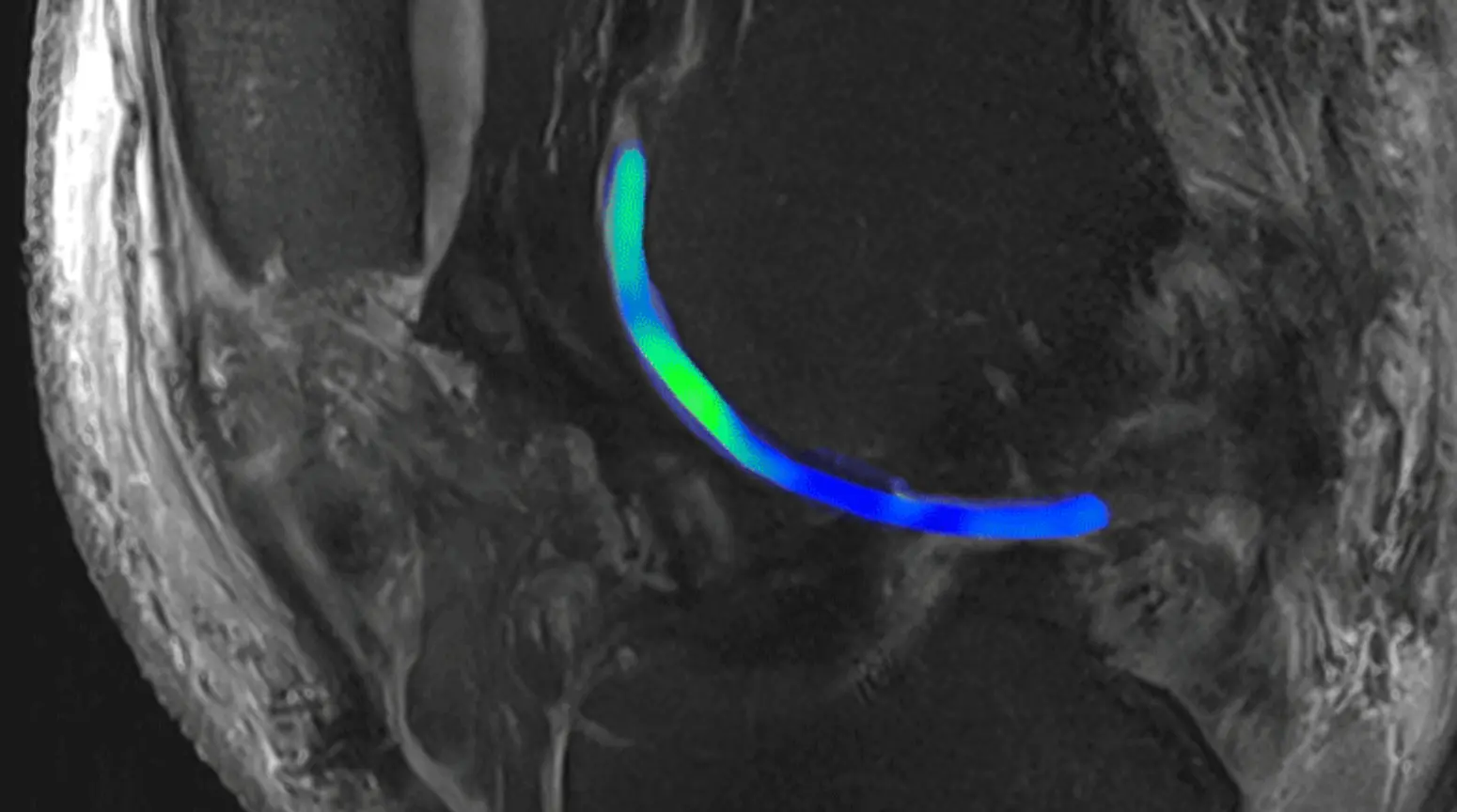
MRI scan of a knee with cartilage highlighted in blue and green.
Traditional MRI uses magnetic pulses to induce a signal from water molecules in the body. This technology differentiates cartilage (blue/light green) from bone. Advanced MRI uses the same approach but applies it to sodium. The technology identifies proteoglycans (bright green), the special ingredient in high-quality cartilage. Regions devoid of this sodium signal point to defective cartilage. Images courtesy Siegfried Trattnig; modified by Fidelis Onwubueke.
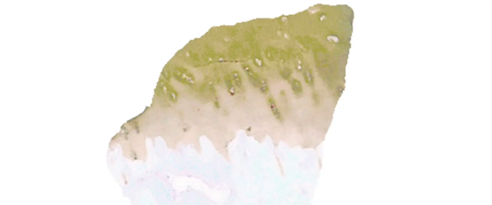
Stained cartilage tissue visualization showing proteoglycans and collagen.
Biopsied cartilage can be stained to reveal the proteins present. Two slivers from the same tissue sample show that proteoglycans and collagen exist together in high-quality cartilage. Proteoglycans appear as a pink stain; collagen appears as a brown stain. Image credit: PhenoPath Laboratories. Animation by Fidelis Onwubueke.

Making the complex clear and engaging.

I produced a cartilage-focused animation and adapted MRI visualizations to help readers understand how researchers assess cartilage quality in regenerative-medicine research.

Capabilities demonstrated

Motion designImage adaptationScientific communicationDrupal publishing
View published project ↗